


Interventional Cardiologists
Interventional cardiologists deal specifically with catheter based treatment (a non-surgical option) of structural heart disease. A catheter is a long flexible tube used to examine and repair damaged or weakened vessels, narrowed arteries and other affected areas of the heart. The cath may be inserted in an artery or vein in your groin, neck or arm and threaded through your blood vessels to your heart.
Through cardiac catheterization (most commonly referred to as a cardiac cath), the cardiologist is able to assess whether there is any disease of the heart muscle, valves or coronary arteries. A cath can also determine the severity of heart problems such as the location and size of plaque deposits and check blood flow and blood pressures in the heart chambers.
A cardiac angiogram is one of the procedures that can be done during a cath. A small amount of a contrast dye is injected into blood vessels using the catheter. The x-ray machine takes a series of images (angiograms) which will show any areas of narrowing and blockages. If blockages are found, the physician may perform several treatments such as placing a stent into the artery to open up the blockage. To view an animation of a coronary angiogram, click here:
https://watchlearnlive.heart.org/index.php?moduleSelect=angiog
In coronary angioplasty, arteries narrowed by atherosclerosis are widened using stents (a tiny wire mesh tube) inserted into a blood vessel via catheterization. The tip of the catheter has a collapsed stent placed over a balloon. When the targeted part of the artery has been reached, the balloon is inflated and the stent expands, resulting in the widening of the artery wall and improving blood flow. The catheter and balloon are then taken out and the stent stays in the artery permanently. The stent will allow improved blood flow to the heart muscle and will relieve chest pain (angina). To view an animation of an angioplasty, click here: https://watchlearnlive.heart.org/?moduleSelect=angiop

(Source: American Heart Association)
Cardiac Catheterization
Cardiac catheterization (cardiac cath or heart cath) is a procedure to examine how well your heart is working. It examines the inside of your heart’s blood vessels using special x-rays called angiograms. Dye visible by x-ray is injected into blood vessels using a thin hollow tube called a catheter that is inserted into a large blood vessel that leads to your heart. The catheter is inserted in your groin, neck or arm and threaded through your blood vessels to your heart.
- A cardiac cath is performed to find out if you have disease of the heart muscle, valves or coronary (heart) arteries.
- During the procedure, the pressure and blood flow in your heart can be measured.
- A contrast dye visible in X-rays is injected through the catheter. X-ray images show the dye as it flows through the heart arteries. This shows if any arteries are blocked.
- The chances that problems will develop during cardiac cath are low.
View an illustration of a cardiac cath here.
- Cardiac catheterization is also used as part of some procedures to treat heart disease. These procedures include:
- Widening a narrow artery with or without a stent (angioplasty)
- Closing holes in the heart and fixing other congenital defects
- Repairing or replacing heart valves
- Opening narrow heart valves (balloon valvuloplasty)
- Treating irregular heart rhythms with ablation
- Closing off part of your heart to prevent blood clots
Some heart disease treatments, such as coronary angioplasty and coronary stenting, are also done using cardiac catheterization. Usually, you’ll be awake during cardiac catheterization but will be given medications to help you relax.
Cardiac Stent
(Source: American Heart Association)
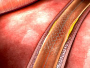 A stent is a small, wire mesh tube that is inserted to permanently prop open a blocked artery in people who are at risk of a heart attack or stroke. When a coronary artery is narrowed by a buildup of fatty deposits called plaque, it can lead to reduced blood flow. If blood flow is reduced to the heart muscle, chest pain can result. If a clot forms and completely blocks the blood flow to part of the heart muscle, a heart attack occurs. Stents help keep coronary arteries open and reduce the chance of a heart attack. Stenting is a common cardiac procedure.
To open a narrow artery, your cardiologist may do a procedure called a percutaneous coronary intervention (PCI) or angioplasty. In it, a balloon-tipped tube (a catheter) is inserted into an artery and moved to the point of blockage then the balloon is inflated. This compresses the plaque and opens the narrowing. When the opening in the vessel has been widened, the balloon is deflated and the catheter is withdrawn. The stent stays in the artery permanently and holds it open. This improves blood flow to the heart muscle and relieves symptoms – usually chest pain.
There are two kinds of stent. One type is covered with drugs that help keep the blood vessel from reclosing. These are called drug-eluting stent. The second type is not coated with drugs and is called a bare metal stent. Your physician will make a determination of which stent is best for you.
Following a stent procedure, you will typically be given an antiplatelet medication to keep platelets from clumping together and forming blood clots in the stent thus blocking the artery.